New organ-on-chip system to help study how neurotoxins move from the gut to the brain
18 December 2024
Researchers have developed a new multiple organ-on-chip system to help study how neurotoxins move from the gut to the brain.
The model system simulated how a neurotoxin in the gut can trigger the brain cell death seen in Parkinson’s disease, in a proof-of-concept study from Hull York Medical School at the University of Hull, the Quadram Institute, the University of Essex and the UK Health Security Agency (UKHSA).
Published in the journal Biomicrofluidics, the study shows how their system recreates the gut-brain axis. This is a bidirectional communication network linking the gastrointestinal tract to the nervous system of the brain, influencing everything from your mood to your digestion, and now believed to be important in the development of Parkinson’s.
The ability to study human-derived cells and tissues of the gut-brain axis, in an interconnected model system, will be a valuable tool to advance our understanding of Parkinsons’ disease and other neurodegenerative conditions.
Research into Parkinsons’ disease is much needed; at least 10 million people globally live with the condition, which attacks and destroys nerve cells in the brain causing tremors, stiffness and other movement-related symptoms. But it also triggers non-motor symptoms elsewhere, including in the gut. Some research indicates gut problems like constipation, and protein accumulations characteristic of Parkinson’s disease brain cell damage, appear in the gut years before a diagnosis of the condition. People with Parkinson’s disease also have changes in their gut microbiome. This has prompted researchers to look at the gut-brain axis.

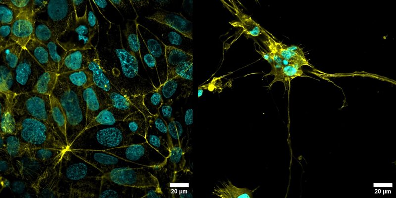
Image of gut cells (left) and brain neuronal cells (right) cultured within the connected MPS devices. Image: Emily Jones and the QIB Advanced Microscopy Facility.
But studying the way these two organ systems interact is difficult. Scientists can grow individual human cells in the laboratory, but these don’t necessarily reflect what happens in the body, due to the complex connectivity networks linking the gut and brain. Research can be carried out in animals to provide more holistic insights, but this raises questions of how well findings translate to humans as well as having ethical concerns.
To fill this gap, miniature microphysiological systems (MPS), have been developed. Also known as “organ-on-chip” technology, MPS allows researchers to grow human cells or tissues in appropriate conditions to mimic how they look, behave and communicate in our bodies.
After receiving a seed start-up grant from the University of Essex, Dr Simon Funnell at the UK Health Security Agency (UKHSA) and Dr Ben Skinner at the University of Essex teamed up with Prof Simon Carding, Dr Emily Jones and Dr Aimee Parker from the Quadram Institute and Prof John Greenman and Dr Lydia Baldwin from the Centre for Biomedicine (Hull York Medical School) at the University of Hull to develop a new and simple-to-use, cost-effective MPS device to mimic the gut-brain axis.
The simplified MPS designed by the University of Hull makes it easier to use without specialist training, and adaptable for use to study a range of disorders. This will help reduce our reliance on the use of animals in this type of research. The model can also be deployed in high-containment laboratory settings, so could be used to track the course of dangerous infections.
Prof John Greenman from the Centre for Biomedicine, Hull York Medical School, at the University of Hull, said: “Each time our devices are used by colleagues to answer different clinical questions we learn how to improve and adapt them in terms of capabilities, robustness and ease of use; we haven’t finished yet.”
Dr Simon Funnell, Group Leader at the Quadram Institute and Scientific Leader at the UK Health Security Agency, said: “Following the protracted illness and death of my mother from Parkinson’s disease, I made a commitment to try to contribute toward innovative research that aims to assist the development of new drugs, therapeutics or treatments to prevent or protect the public from the threat of early onset neurological decline” said
“This collaborative research has applications outside of Parkinson’s disease and UKHSA is working to use this tool to better understand the impact of infectious diseases on the body and evaluate treatments and vaccines.”
To create the gut-brain MPS, two devices are combined in series; connected via tubing representing the blood flow. In the first device, a layer of cells representing the gut lining form a selectively permeable barrier between the contents of the gut and the rest of the body. In the second connected device, the researchers cultured human-derived brain neuronal cells of a type known to be susceptible to neurotoxins.
Dr Ben Skinner from the University of Essex said: “This is a very exciting development and really shows the power of innovation and collaboration between scientists from across the country.
“This project was sparked after a chance meeting between Dr Funnell and I at a University of Essex lab challenge day and it is incredible to see how it has developed. The paper has been the culmination of more than five year’s work, and I hope this technology can make a real difference in fighting Parkinson’s disease.”
This means the MPS can simulate not just how neurotoxins cross the gut lining, but also how they travel to the brain and interact with cells there. In this proof-of-concept study, the neurotoxin was introduced into the gut, and was seen to kill off the brain cells, without affecting the cells of the gut lining as it passed across them.
Dr Emily Jones from the Quadram Institute said: “We hope this new multi-organ MPS will provide a valuable tool for unravelling the complex interactions between the gut and brain.
“By allowing us to study human-derived cells in an interconnected model, we aim to gain deeper insights into disease mechanisms and potentially identify new therapeutic targets that can protect against neuronal inflammation and cell death. This approach could revolutionise our understanding of neurological disorders, paving the way for more effective treatments and ultimately improving the lives of millions affected by these conditions.”
Professor Isabel Oliver, Chief Scientific Officer at the UK Health Security Agency, said: “We are using Organ on Chip technology to better understand the impact of viruses on the human body, allowing us to evaluate and predict the effectiveness of vaccines and treatments.
“We’ve already developed this technique to look at the impact of COVID-19 on the lungs and we are now working to expand this tool to study other organs and how they are impacted by COVID-19 and other infections.”
The research was funded by the Economic and Social Research Council (UKHSA and University of Essex, Public Health Challenge Lab) and Biotechnology and Biological Sciences Research Council (QIB), both part of UKRI.
For more information please email pr@hull.ac.uk or call 07484 534322.
Image: Emily Jones and the QIB Advanced Microscopy Facility
Reference: Emily J. Jones, Benjamin M. Skinner, Aimee Parker, Lydia R. Baldwin, John Greenman, Simon R. Carding, Simon G. P. Funnell; An in vitro multi-organ microphysiological system (MPS) to investigate the gut-to-brain translocation of neurotoxins. Biomicrofluidics 1 September 2024; 18 (5): 054105.